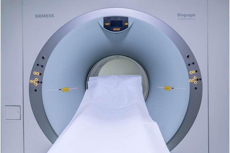
At the intersection of AI and clinical science, there might be rising hobby within the utilization of machine studying to present a enhance to imaging files captured by magnetic resonance imaging (MRI) technology. Recent stories repeat that extremely-excessive-self-discipline MRI at 7 Tesla (7T) could more than seemingly own far better resolution and clinical advantages over excessive-self-discipline MRI at 3T in delineating anatomical structures that are famous for figuring out and monitoring pathological tissue, in particular within the brain.
Most clinical MRI tests within the U.S. are performed with 1.5T or 3T MRI programs. As goal now not too long within the past as 2022, the National Institutes of Health documented handiest about 100 7T MRI machines being worn for diagnostic imaging worldwide.
Researchers from UC San Francisco developed a machine studying algorithm to present a enhance to 3T MRIs by synthesizing 7T-esteem photos that approximate right 7T MRIs. Their mannequin enhanced pathological tissue with extra fidelity for clinical insights and represents a new step towards evaluating clinical capabilities of synthetic 7T MRI objects.
The look became presented Oct. 7 at the 27th International Convention on Clinical Image Computing and Computer Assisted Intervention (MICCAI).
“Our paper introduces a machine-learning model to synthesize high-quality MRIs from lower-quality images. We demonstrate how this AI system improves the visualization and identification of brain abnormalities captured by MRIs in Traumatic Brain Injury,” acknowledged senior look author Reza Abbasi-Asl, Ph.D., UCSF Assistant Professor of Neurology.
“Our findings highlight the promise of AI and machine learning to improve the quality of medical images captured by less advanced imaging systems.”
Greater to behold TBI and multiple sclerosis with
UCSF researchers serene imaging files from sufferers recognized with relaxed anxious brain damage (TBI) at UCSF. They designed and professional three neural community objects to present image enhancement and 3D image segmentation the utilization of the generated artificial-7T MRIs from the customary 3T MRIs.
The photos generated with the brand new objects supplied enhanced pathological tissue for sufferers with relaxed TBI. They selected an instance space with white topic lesions and microbleeds in subcortical areas to make employ of for comparison. They found pathological tissue became less complicated to behold in synthesized 7T photos. This became evident within the separation of adjoining lesions and the sharper contours of subcortical microbleeds.
Additionally, the synthesized 7T photos better captured the diverse sides internal white topic lesions. These observations moreover highlight the promise of the utilization of this technology to toughen diagnostic accuracy in neurodegenerative problems similar to multiple sclerosis.
Whereas synthetization suggestions primarily based totally on machine studying frameworks repeat unprecedented performance, their application in clinical settings will require huge validation. The researchers say that future work must encompass huge clinical overview of the mannequin findings, clinical score of mannequin-generated photos, and quantification of uncertainties within the mannequin.
More files: Qiming Cui et al, 7T MRI Synthesization from 3T Acquisitions, Clinical Image Computing and Computer Assisted Intervention – MICCAI 2024 (2024). DOI: 10.1007/978-3-031-72104-5_4
Citation: Bettering MRI with AI to toughen prognosis of brain problems (2024, October 14) retrieved 8 December 2024 from https://medicalxpress.com/files/2024-10-mri-ai-prognosis-brain-problems.html
This file is area to copyright. Other than any stunning dealing for the motive of private look or analysis, no phase will be reproduced without the written permission. The disclose material is supplied for files capabilities handiest.


